By Avani V
Volunteer
Introduction:
Forensic anthropology is the application of the anatomical science of anthropology and its various subfields, including forensic archeology and forensic taphonomy in a legal setting. Sexual dimorphism is the variation represents both genetic and environmental factors which impact methodologies used to estimate sex from human skeletal remains.
Factors that contribute to sexual dimorphism are sex hormones, Genetics and inflammation. Examples of sexual dimorphism in skeletons are pelvis, cranium, long bones, vertebral bodies.
Sexual dimorphism in the development of the skeleton and in the incidence of skeletal diseases is well described. In general females, at any given age, have a lower bone mass than males. In addition, women predominate in the incidence of osteoporosis while men more frequently develop Paget’s disease of bone. The organization of bone into a functional skeleton, which provides organisms with structural integrity, is the net result of the activity of osteoclasts, which resorb bone, osteoblasts, which form bone and osteocytes, which coordinate the activities of the other two cell types
History: The study of sexual dimorphism in skeletons has been on going for decades with research involving variety of techniques and populations. “António Mendes Correia” in the early 20th century, Correia documented sexual differences in the skeleton, including the shoulder girdle, vertebral column, and arm. Principal Component Analysis (PCA) A PCA was performed on the cranial form of known sex individuals from different populations.
Fig: Skeletal Remains Of Roman Aristocrat
Sexual Dimorphism In Skeletal Remains:
Sex Determination From Skull:
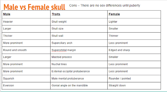
Supraorbital ridge: Males tend to have a more prominent and thicker brow ridge compared to females, which appears as a pronounced ridge above the eye sockets.
Forehead slope: A male skull typically has a more sloping forehead, while a female skull has a more vertical and rounded forehead.
Chin shape: Male jawlines tend to be more square and robust, with a larger chin, whereas female chins are usually more pointed.
Orbital shape: Female eye sockets tend to be more rounded, while male eye sockets can appear more rectangular
Fig: Tabulation of Determination of Sex from skull
Fig: Labelled Parts Of Skull
Sex Determination From Pelvis:
The pelvis is one of the most sexually dimorphic regions of the skeleton. The overall shape of the male pelvis is narrow and steep, and the female pelvis is generally broader, with a larger pelvic outlet to facilitate pregnancy and childbirth. Since this is the only region of the skeleton where there is such a key functional difference, the pelvis proves to be one of the most important bone for determining biological sex. The accuracy of sex determinations from the skeleton based on the pelvis alone is usually placed at around 96%, but this will vary between populations as well as the degree of preservation and fragmentation.
Fig: Tabulation of Determination Of Sex From Pelvis
Fig: Male And Female Pelvis
Sex Determination From Long Bones:
shaft of long bones are relatively rough and the particular surfaces and ends larger in males and in females shaft of the bones are relatively smooth and the articular surfaces and ends smaller.
Fig: Long Bone Measurements
Conclusion:
Analyzing skeletal remains allows for a reliable assessment of an individual’s sex based on distinct morphological features ,with pelvis being the most sexually dimorphic region providing the most accurate determination other bones like skull and long bones can also provide information when considering size ,shape and muscle attachment points. Although individual variation and population differences should always be taken in to picture when interpreting sexual dimorphism.
References:
Thank You


0 Comments